Distal radius fractures are one of the most common orthopedic fractures. They can occur from a low energy injury (i.e. a fall on an outstretched hand is the most common mechanism) or high energy (i.e. motor vehicle accident) injury. Often there will be a visible deformity of the wrist due to bony displacement.
The evaluating provider will order Xrays if there is a suspicion of a fracture. If the fracture extends into the joint, a CT scan may also be ordered to further evaluate the fracture to determine the best treatment option. The distal radius can tolerate a certain amount of angulation and displacement. However, there is less tolerance for displacement of the joint fragments. Your orthopedic team will know how to determine the best treatment option for your specific injury pattern.

If there is acceptable alignment of the fracture, then the wrist may be splinted for a few days then casted. X-rays will be taken a week or two later to ensure the fracture has not shifted and that alignment remains acceptable. Usually with non-operative treatment, the cast is left in place for 6 weeks at which time X-rays are taken to assess the amount of bony healing. If adequate healing, then the patient is transferred to a removable brace and therapy is often initiated to begin restoring motion. Full return to sport/activity will be determined by the orthopedic team but usually occurs at about 8-12 weeks depending on the desired sport/activity and level of bone healing.
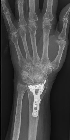
If the injury warrants surgery, there are a few different techniques to provide adequate fixation and the best chance for healing. The most common surgical approach is performing an open reduction and internal fixation (ORIF) using a volar approach and plate and screw placement (see bottom picture). The benefits of surgical fixation is getting an excellent reduction, solid fixation and initiating early motion and reducing the risk of prolonged stiffness in the wrist.
After surgery, a plaster splint will be placed and left in place until the initial postoperative visit at roughly two weeks. At this time, the splint will be removed, X-rays will be taken and the patient will be transitioned into a removable wrist brace and gentle therapy will be initiated to prevent further stiffness. The following protocol may be modified according to the quality of the bone and the fracture pattern. The goals of rehabilitation are to limit swelling, maintain finger motion, and gradually regain wrist range of motion and strength while protecting the fracture as it heals.