Background
The hip is a ball and socket joint comprised of the femoral head (ball) and the acetabulum of the pelvis (socket). This joint is prone to high levels of stress, bearing the most weight of any other joint, secondary to the knee. The femoral head is attached to the pelvis via a fibrous synovial lined joint capsule. Additional stability and function is provided by multiple surrounding ligaments, tendons and muscles. Overlying the femoral head and lining the acetabulum is a smooth and tough covering of hyaline cartilage. This cartilage is important in providing protection to the bony surfaces of the hip joint and allowing for a smooth pain free range of motion. If either of the cartilage surfaces degenerates over time, pain can occur. This degenerative process is multi-factorial in its cause and includes both modifiable and non-modifiable risk factors.
Some modifiable risk factors include prior trauma involving the articular surfaces of the joint, obesity, muscle weakness, and chronic heavy loading of the joint. Non-modifiable risk factors include gender (females > males), age, genetics and developmental hip abnormalities (dysplasia, Legg-Calve-Perthes disease, slipped capital femoral epiphysis).
Diagnosis
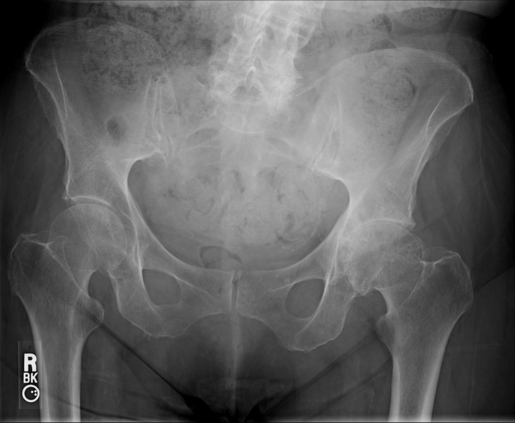
The diagnosis of hip arthritis can usually be obtained through a detailed history, physical exam and X-rays. The patient's history may include symptoms of groin pain, pain "deep in the joint", pain in a "c-clamp" distribution around the hip, thigh and even occasional knee pain. Physical exam may show limitations in range of motion and certain provocative measures can elicit pain in the groin. X-rays will show decreased joint space and even surrounding osteophytes from the femoral head or acetabular rim. On occasion, an MRI may be needed to confirm cartilage degeneration.
Treatment
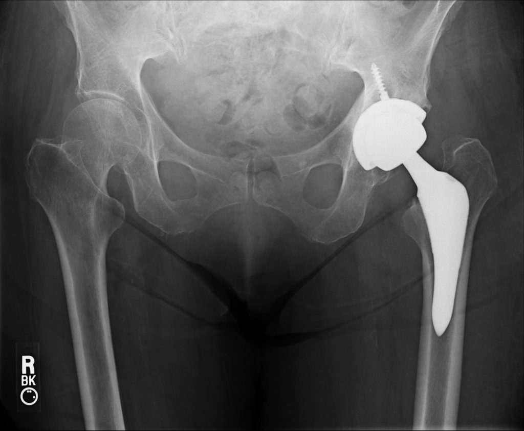
In the early arthritic hip, treatment may initially consist of avoidance of aggravating factors and over the counter medications such as acetaminophen and/or NSAIDs. As the osteoarthritis worsens, intra-articular steroid injections may be offered. These injections can be performed in our office under ultrasound guidance, avoiding the need to have this done under X-ray at a hospital. As a result, the cost is much less and the outcome is just as effective. When conservative measures are no longer providing adequate pain relief, the surgical option of total hip replacement may be explored.
There are a few different methods and approaches when performing a total hip replacement. Much of the differences lie in the manner in which the hip joint is accessed. Generally speaking, the three approaches are the anterior, anterior-lateral, and posterior approaches. Although more technically demanding, the direct anterior approach is chosen by Dr. Lee for several reasons. For example, in a direct anterior approach, the muscles around the hip are merely moved out of the way during the surgery. No glute tendons are cut as with a posterior approach. Hip retractors are carefully placed to provide adequate visualization. As a result, no subsequent tendon repair is required, thus, alleviating the need for protecting a repair.
With a posterior approach, tendons around the hip must be cut and pulled out of the way in order to access the joint. These tendons are subsequently repaired but require several months of rehabilitation and healing time. Also, the posterior capsule of the hip joint must be opened to access the hip which puts the patient at increased risk for posterior hip dislocations. Therefore, after a posterior total hip, the patient requires several range of motion limitations including avoidance of deep hip flexion and crossing your legs. Therapy is also required to strengthen the surrounding muscles which can take around 3 months before the hip precautions are removed.