Meniscus Injuries
Background
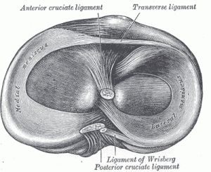
 The meniscus is a specialized cartilage found in the knee that lies between the femur and the tibia. The primary purpose of the meniscus is to decrease the contact pressure between the femur and the tibia. The secondary purpose of the meniscus is to contribute to the stability of the knee. Determining if a patient has a meniscus tear is based on the physical examination and imaging – typically a knee MRI.
The meniscus has a unique anatomy with an overall shape similar to the letter C with robust collagen fibers running the length of the meniscus. These fibers are strongly anchored both anteriorly and posteriorly and these anchor points otherwise known as roots are critical to the meniscus function. In cross section it has a triangular shape in cross section with the blood supply coming from the peripheral edge. As the meniscus tapers centrally it has essentially blood supply
Injuries

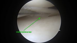
Meniscus injuries are very common and arthroscopic partial meniscus resection is the most common surgery performed by orthopedic surgeons each year. The meniscus can be injured by an acute traumatic event or by a degenerative process. In the younger population, meniscus injuries are related to large shifts in the knee such as when a ligament is torn. In the elderly population, the meniscus tissue is less pliable and subject to tearing with more minor trauma. If a knee becomes arthritic the irregularity of the cartilage or exposed bone can pinch/grind the meniscus tissue and cause tearing.
There is a great degree of variability in the type of meniscus tears. The basic tear patterns are horizontal tears, longitudinal tears, radial tears and root tears. How to approach these tears surgically depends upon the size of the tear, the blood supply to the tear, overall health of the joint, and the age of the patient.
Treatment

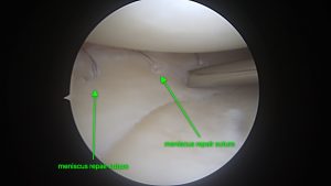
Treatment options include injection therapy with either biologics or steroid, partial resection, repair of the meniscus and in select cases meniscus transplant. A partial resection of the meniscus is the most common orthopedic procedure done each year. A partial resection means that the torn portions of the meniscus are removed and at times this is the best option for the knee. However, the meniscus has a very important function of distributing the weight of the body across the knee so the cartilage does not experience as much pressure. In my hands, I am aggressive about repairing the meniscus when I can to preserve the long-term health of the knee. When a meniscus is unsalvageable and the underlying knee cartilage is in good condition, a meniscus transplantation can be performed – restoring more normal knee function.
Rehabilitation
Depending upon the surgery perform we may or may not put restrictions in regards to knee motion and weight bearing. As a general rule, if a menisectomy is performed, full range of motion of the knee is allowed as well as full weight bearing. However in cases of meniscus repair or transplantation, there is typically a 4-6 week restriction on motion and weight bearing.
For more information regarding meniscus surgery rehabilitation, please revere to our rehabilitation protocols.
MENISCUS REPAIR PROTOCOL
This protocol is intended to be a framework for the rehabilitation of patients who have undergone meniscus repair or meniscus root repair. Some personalized changes are possible. The intent of this protocol is to provide the clinician with a guideline of the post-operative rehabilitation course. If a clinician requires assistance in the progression of a post-operative patient they should consult with the referring Surgeon or PA.
Progression is usually determined by clinical findings that meet certain criteria.
Key Factors in determining progression of rehabilitation after Meniscal repair include:
- Anatomic site of tear
- Suture fixation (failure can be caused by too vigorous rehabilitation)
- Location of tear (anterior or posterior)
- Other pathology (ligamentous injury)
Phase I – Weeks 1-6:
Goals:
- Diminish inflammation and swelling
- Restore limited ROM
- Reestablish quadriceps muscle activity
Stage 1: Immediate Postoperative Day 1- Week 3
- Ice, compression, elevation
- Electrical muscle stimulation
- Brace locked at 0 degrees when ambulating (as applicable)
- ROM 0-90
Motion is limited for the first 7-21 days, depending on the development of scar tissue around the repair site. Gradual increase in flexion ROM is based on assessment of pain and site of repair (0-90 degrees).
- Patellar mobilization
- Scar tissue mobilization
- Passive ROM
- Exercises
- Quadriceps isometrics
- Hamstring isometrics (if posterior horn repair, no hamstring exercises for 6 weeks)
- Hip abduction and adduction
- Flat-foot weight bear at this time with crutches and brace at 0-90 degrees.
Stage 2: Weeks 4-6
- Depending on repair, limited weight bearing may begin at this time with brace locked in extension.
- Progressive resistance exercises (PREs) 1-5 pounds.
- Limited range knee extension (in range less likely to impinge or pull on repair)
- Toe raises
- Cycling (no resistance)
- PNF with resistance
- Unloaded flexibility exercises
- Proprioception training with brace locked at 0 degrees
Phase II: Moderate Protection- Weeks 6-10
Criteria for progression to phase II:
- ROM 0-90 degrees
- No change in pain or effusion
- Good Quadriceps control
Goals:
- Increased strength, power, endurance
- Normalize ROM of knee
- Prepare patients for advanced exercises
Exercises:
- Strength- PRE progression
- Flexibility exercises
- Lateral step-ups
- Mini-squats
Endurance Program:
- Swimming (no frog kick), pool running- if available
- Cycling
- Stair machine
Coordination Program:
- Balance board
- Pool sprinting- if pool available
- Backward walking
- Plyometrics
Phase III: Advanced Phase- Weeks 11-15
Criteria for progression to phase III:
- Full, pain free ROM
- No pain or tenderness
- Satisfactory clinical examination
- SLR without lag
- Gait without device, brace unlocked
Goals:
- Increase power and endurance
- Emphasize return to skill activities
- Prepare for return to full unrestricted activities
Exercises:
- Continue all exercises
- Increase plyometrics, pool program
- Initiate running program
Return to Activity: Criteria
- Full, pain free ROM
- Satisfactory clinical examination
Criteria for discharge from skilled therapy:
1) Non-antalgic gait
2) Pain free /full ROM
3) LE strength at least 4/5
4) Independent with home program
5) Normal age appropriate balance and proprioception
6) Resolved palpable edema
PARTIAL MENISCECTOMY
A partial medial or lateral meniscectomy or loose body removal is performed in order to decrease pain in the knee associated with constant irritation within the knee joint. After surgery the knee may continue to be sore for some time. Patients may also continue to have pain in the anterior of the knee especially during kneeling for 4-6 months following arthroscopic surgery. Rehabilitation after meniscectomy may progress aggressively because there is no anatomic structure that requires protection. Progression to the next phase is based on clinical criteria and meeting the established goals for each phase.
Phase I – Acute Phase:
Goals:
- Diminish pain, decrease swelling
- Restore knee range of motion (goal 0-115, minimum of 0 degrees extension to 90
degrees of flexion to progress to phase II)
- Reestablish quadriceps muscle activity/re-education (goal of no quad lag during
straight leg raise.)
- Patients may require education regarding weight bearing as tolerated, use of crutches, icing, elevation and the rehabilitation process
Weight bearing:
- Weight bearing as tolerated. Patients may use two crutches initially and quickly progress to one crutch and then off of crutches as swelling, pain, and quadriceps function dictates.
Modalities:
- Cryotherapy 20 min on and 20 min off as tolerated for the first few days, then as needed throughout the day, this may be accomplished with ice packs or a Thermotec device.
- Electrical stimulation to quadriceps for functional retraining as appropriate
- Manual therapy as needed for fluid mobilization.
Therapeutic Exercise:
- Quadriceps sets
- Straight leg raises
- Hip adduction, abduction and extension
- Calf sets (ankle pumps)
- Gluteal sets
- Heel slides
- Squats to 60 degrees
- Active-assisted ROM stretching, emphasizing full knee extension (flexion to tolerance)
- Hamstring and gastroc/ soleus and quadriceps stretches
- Bicycle for ROM when patient has sufficient knee ROM. May begin partial revolutions to recover motion if the patient does not have sufficient knee flexion
Phase II: Internal Phase :
Goals:
- Restore and improve muscular strength and endurance
- Reestablish full pain free ROM
- Gradual return to functional activities
- Restore normal gait without an assistive device
- Improve balance and proprioception
Weight bearing status:
Patients may progress to full weight bearing as tolerated without antalgia. Patients may
require one crutch or cane to normalize gait before ambulating without assistive device.
Therapeutic exercise:
- Continue all exercises as needed from phase one
- Toe raises- calf raises
- Hamstring curls
- Continue bike for motion and endurance
- Cardio equipment- stairmaster, elliptical trainer, treadmill and bike as tolerated.
- Lunges- lateral and front
- Leg press
- Lateral step ups, step downs, and front step ups
- Knee extension 90-40 degrees
- Closed kinetic chain exercise terminal knee extension
- Four way hip exercise in standing
- Proprioceptive and balance training
- Stretching exercises- as above, may need to add ITB and/or hip flexor stretches
Phase III – Advanced activity phase:
Goals:
- Enhance muscular strength and endurance
- Maintain full ROM
- Return to sport/functional activities/work tasks
Therapeutic Exercise:
- Continue to emphasize closed-kinetic chain exercises
- May begin plyometrics/ vertical jumping
- Begin running program and agility drills (walk-jog) progression
- Sport specific drills
Criteria for discharge from skilled therapy:
1) Non-antalgic gait, pain free /full ROM
2) LE strength at least 4+/5
3) Independent with home program
5) Normal age appropriate balance and proprioception
6) Resolved palpable edema
A partial medial or lateral meniscectomy or loose body removal is performed in order to decrease pain in the knee associated with constant irritation within the knee joint. After surgery the knee may continue to be sore for some time. Patients may also continue to have pain in the anterior of the knee especially during kneeling for 4-6 months following arthroscopic surgery. Rehabilitation after meniscectomy may progress aggressively because there is no anatomic structure that requires protection. Progression to the next phase is based on clinical criteria and meeting the established goals for each phase.
